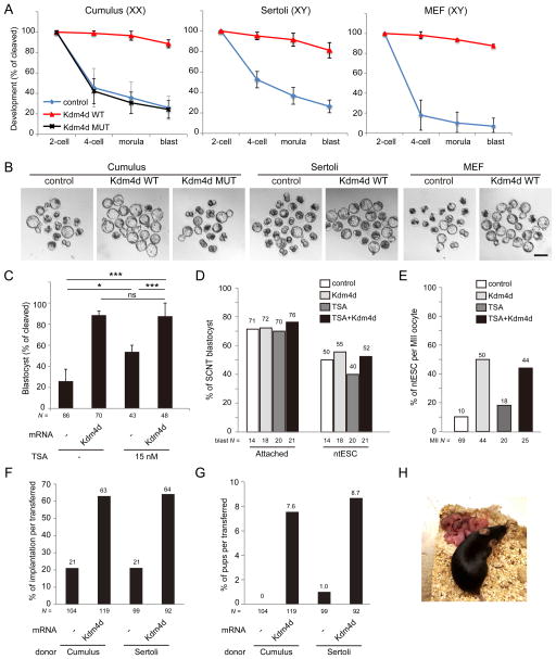
Figure 4. Injection of Kdm4d mRNA improves developmental potential of SCNT embryos.
(A) Kdm4d mRNA injection greatly improves preimplantation development of SCNT embryos derived from cumulus cells, Sertoli cells and MEF cells. Shown is the percentage of embryos that reaches the indicated stages. XX and XY indicate the sex of donor mice. Error bars indicate s.d.
(B) Representative images of SCNT embryos after 120 hours of culturing in vitro. Scale bar, 100 μm.
(C) Kdm4d mRNA injection has additional effect over the treatment with Trichostatin A (TSA; 15 nM). Shown is the percentage of embryos that reached the blastocyst stage at 96 hpa. * P < 0.05, ** P < 0.01, *** P < 0.001. ns, not significant.
(D) Bar graph showing the efficiency of attachment to the feeder cells and ntESC derivation of SCNT blastocysts.
(E) Bar graph showing the efficiency of ntESC derivation. The efficiency was calculated based on the total number of MII oocytes used for the generation of SCNT embryos.
(F, G) Implantation rate (F) and birth rate (G) of SCNT embryos examined by caesarian section on E19.5.
(H) An image of an adult female mouse derived by SCNT of a cumulus cell with Kdm4d mRNA injection and its pups generated through natural mating with a wild-type male.
See also Tables S1–3.

