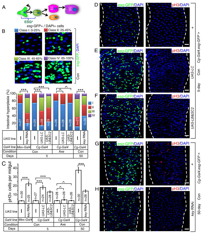
Figure 2. Systemic inflammation caused by old fat body results in midgut hyperplasia.
A. An intestinal stem cell (ISC) produces a new ISC and an enteroblast (EB). EBs differentiate into enteroendocrine cells (EE) or premature enterocytes (red). ECs further differentiate to mature polyploid ECs (purple). Both ISCs and EBs express ESCARGOT (ESG+).
B. Midgut hyperplasia based on esg-GFP in P1-4 region. The number of esg-GFP+ cells counted was divided by total cells counted based on DAPI staining. The percent of esg-GFP+ cell (% of intestinal hyperplasia) was grouped into four classes and plotted (bottom). ^p>0.05, **p<0.01, ***p<0.001, Wilcoxon two-sample test.
C. Midgut hyperplasia based on pH3 staining in the whole midgut. The average pH3+ cells/midgut was plotted. Error bars, SEM. ^p>0.05, ***p<0.001, Student’s t-tests.
D–H. Expression of PGRP-LC (LC, E) or PGRP-SC2 plus PGRP-LB (SC2&LB, F) in young fat bodies increased midgut esg-GFP+ (green) and pH3+ (red) cells compared to that of the control (D). Depletion of KEY in the fat body by RNAi (H) reduced the number of midgut esg-GFP+ and pH3+ cells in old midguts compared to that of control (G). DAPI (blue), nuclei. Scale bars, 20 μm.
Con and Axe, conventional and axenic conditions. n, numbers of midguts analyzed.
See also Figure S2.

