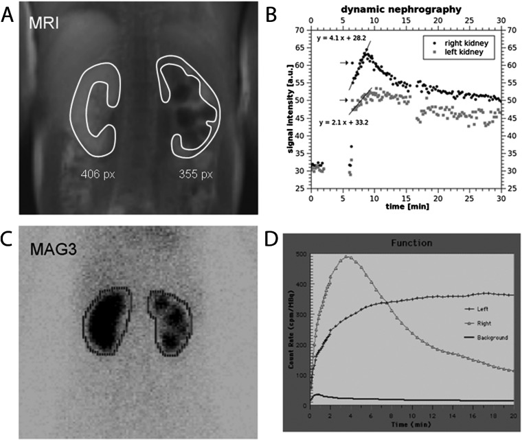
Figure 1.
MR nephrography of an 11-year-old girl with scarring of the left kidney owing to recurrent pyelonephritis associated with reflux. (a) Upper left image shows a TurboFLASH image from the dynamic series acquired in the parenchymal phase after contrast medium injection. Typical region of interest (ROI) definition is indicated (white lines). (b) The upper right image displays signal-intensity–time curves of both kidneys. Split renal function is calculated from the best-fit linear regression line to the signal increase in the parenchymal phase weighted with the size of the ROI. Lower images show the corresponding mercaptoacetyltriglycine (MAG3) examination [(c) ROI definition, (d) corresponding intensity curves]. MR nephrography resulted in split renal function of 69% (right side): 31% (left side), MAG3 scintigraphy 67%:33%.

