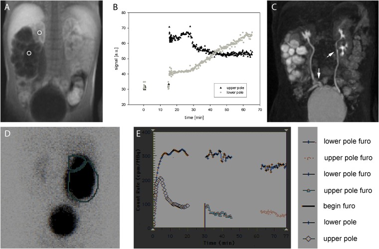
Figure 2.
Upper row: (a) MR urography of a 2-year-old boy with double collecting system on both sides and ureteropelvic junction obstruction of the right lower pole. Two regions of interest (ROIs) were placed in the right upper and lower pole, respectively. (b) Corresponding signal-intensity–time curves: right upper pole shows normal signal behaviour with a fast increase of signal intensity after bolus injection to a plateau phase, which is owing to constant filtration of contrast medium into the urine without obstruction. After 20 min, furosemide (Furo) is applied resulting in the decline of the plateau to a lower level caused by reduced water reabsorption during constant filtration. Right lower pole exhibits a clearly different behaviour; a constant increase of signal intensity is displayed, which does not decline after furosemide application indicating functionally relevant obstruction of urinary transport. (c) Maximum-intensity projection of the three-dimensional data set acquired approximately 10 min after bolus injection during a short pause of the dynamic acquisition. Ureter fissus on both sides is indicated with arrows. Lower row shows corresponding mercaptoacetyltriglycine (MAG3) scintigraphy: (d) ROI definitions and (e) corresponding intensity curves.

