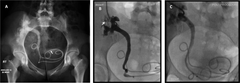
Figure 4.
(a) An 18-year-old female live-related renal transplant recipient. Intravenous urogram demonstrates duplex renal transplant with delayed excretion from the upper moiety and a standing column within the ureter (routline arrow). The upper moiety ureteric stent has migrated into the bladder (solid arrow). Normal appearing calyces are seen in relation to the lower moiety, and the ureteric stent to this section of the duplex kidney appears to be in a satisfactory position. There is contrast seen in the bladder, which is likely to represent drainage from the lower moiety. (b) Nephrostogram performed 2 days following nephrostomy placement within the dilated upper moiety shows extravasation from calyces in the upper moiety of the transplant duplex kidney (arrow), in addition to a dilated upper moiety ureter to the level of the distal ureter. (c) 8.5-French, 12-cm ureteric stent placed with the upper moiety ureter in satisfactory position. A temporary covering nephrostomy was also placed.

