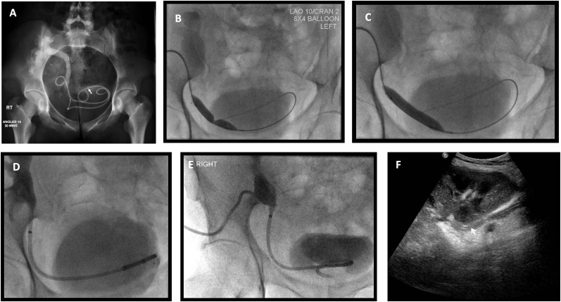
Figure 5.
Management of the case presented in Figure 2. (a) Nephrostogram following nephrostomy placement indicates tight distal ureteric stricture (arrow). (b) The stricture was crossed with a hydrophilic Terumo™ wire (Terumo Corporation, Somerset, NJ) and dilated with an 8 × 40 -mm balloon. Waisting of the balloon confirms the presence of a stricture. (c) Abolishment of the balloon waist indicating satisfactory dilation of the stricture. (d) An 8.5-French antegrade double-J stent is sited and a locked covering nephrostomy left in situ. (e) Following contrast administration via the indwelling nephrostomy, there is prompt drainage into the urinary bladder. The nephrostomy was consequently removed under fluorosocopy over a standard wire. (f) Ultrasound performed 7 days following nephrostomy removal demonstrated satisfactory appearance of the transplant kidney with no residual hydronephrosis and ureteric stent in situ (arrow).

