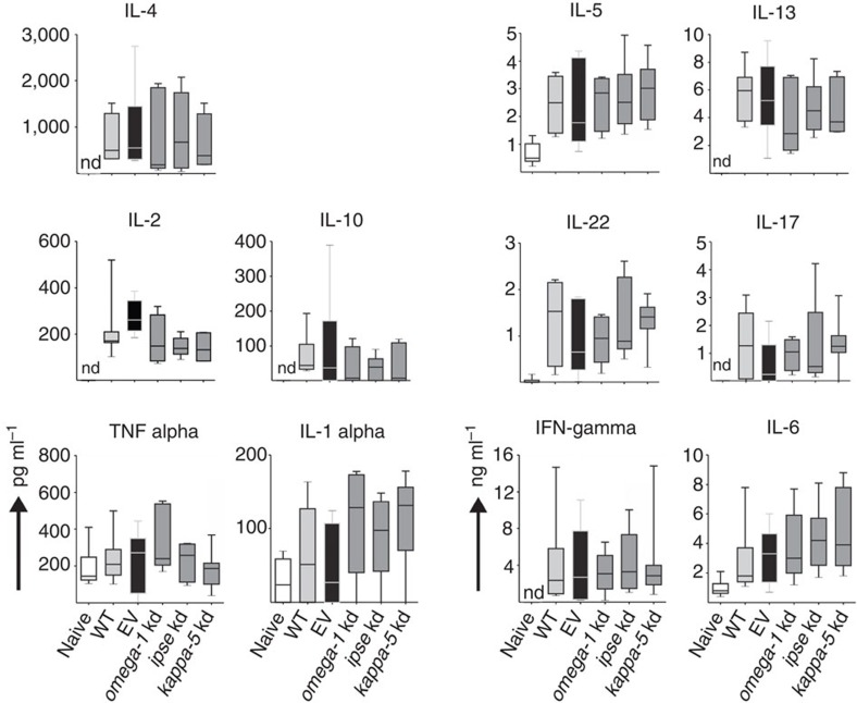
Figure 4. Ex vivo cytokine production of lung cell suspensions after antigen-specific restimulation.
Lung cells of BALB/c mice were analysed for cytokine secretion 15 days after injection, via the tail vein, with PBS (naive), eggs of S. mansoni treated with no virus (WT), empty-vector (EV), or lentiviruses containing shRNAmirs directed at omega-1, ipse or kappa-5 transcripts. Lung cell suspensions were restimulated with SEA (20 μg ml−1) for 72 h and culture supernatants (n=5–7) assayed by cytometric bead array. Box plot representing the median and upper/lower quartile. Whiskers indicate the highest or lowest value. IL, interleukin; IFN, interferon; kd, knockdown; nd, not detected.

