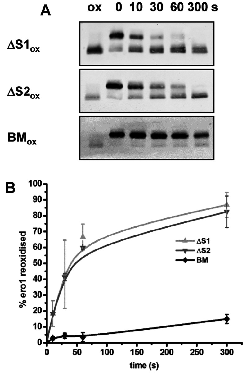
Figure 4. Both PDI active sites contribute to Ero1α re-oxidation and inactivation, as does the PDI b′-binding domain.
Reduced Ero1α (2 μM) was incubated under anaerobic conditions with the oxidized (ox) PDI mutants ΔS1 or ΔS2 or the PDI-binding mutant (BM) all at 10 μM. (A) After the time points indicated (seconds) the redox state of Ero1α was frozen with NEM and samples analysed by non-reducing SDS/PAGE and Western blotting with an antibody against Ero1α. (B) The fraction of Ero1α re-oxidized for three independent experiments was quantified and the mean value calculated and plotted against the time of incubation (error bars represent S.D.).

