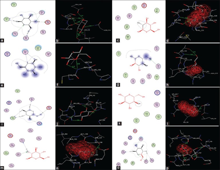
Fig. 5.

Molecular docking of glucose in to S. aureus glk A and other bacterial glk A active sites
(a) Two-dimensional (2D) linear representation of glucose interaction in the S. aureus glk A active site. (b) Three-dimensional (3D) graphical representation of glucose interaction in the active site of S. aureus glk A. (c, d) Clostridium difficile glk A. (e, f) Bacillus anthracis glk A. (g, h) Bacillus subtilis glk A. (i, j) Renibacterium salmoninarum glk A. (k, l) Streptococcus pneumonia glk A. (m, n) Escherichia coli glk A and (o, p) Enterococcus faecalis glk A.
