
Figure 1.
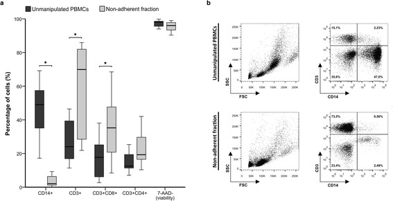
Phenotypic analysis of cells before and after the adherence process. Plastic adherence method was applied to PBMCs from G-CSF mobilized donors (n = 11) and CD14, CD3, CD8, CD4 and 7-ADD expression of the unmanipulated PBMCs and the non-adherent cells were analyzed by flow cytometry (a). Representative figure of the products before and after the adherent process (b). Cells are presented from the CD45+ cell gate.