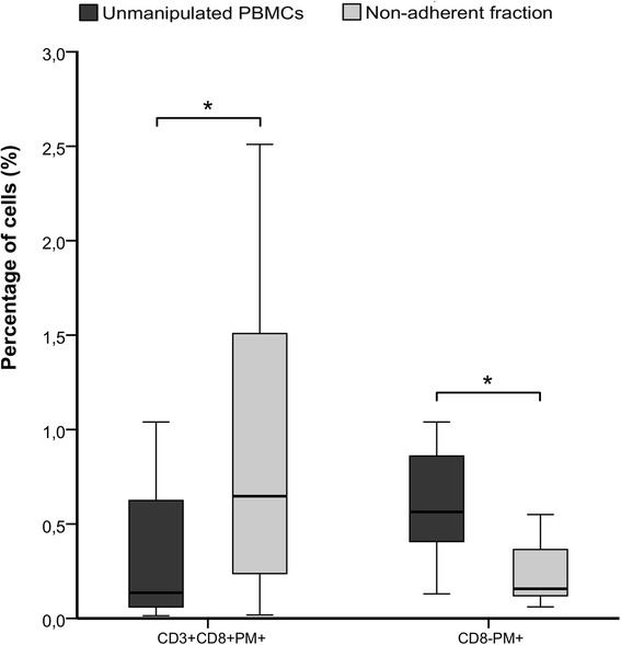
Figure 2.

PM staining before and after the adherence process. Plastic adherence method was applied to PBMCs from G-CSF mobilized donors (n = 11) and PM+ cells were quantified in unmanipulated PBMCs and the non-adherent cell fraction. Specific (CD3+CD8+PM+) and non-specific (CD8-PM+) PM binding was analyzed. Percentages were analyzed from CD45+ cell gate.
