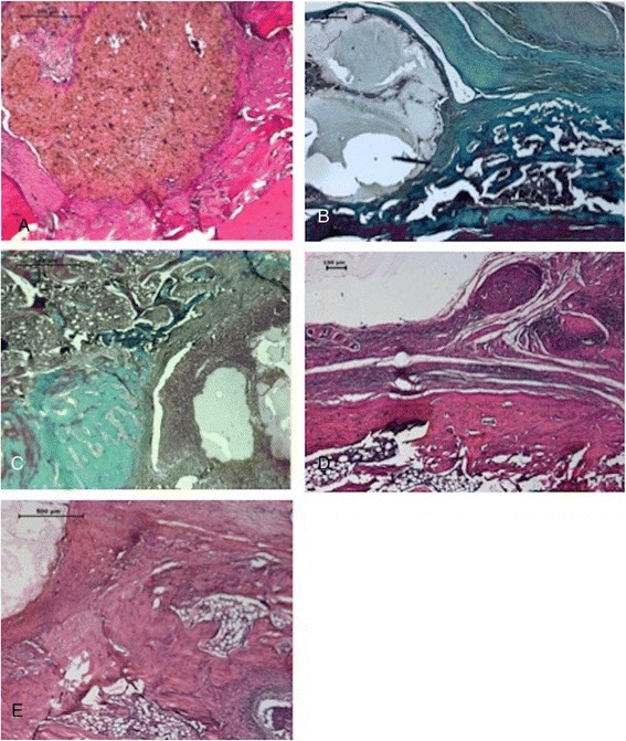
Figure 4.

Histological images (A-E). PLLA/β-TCP without IRO group had a mild tissue reaction in (A) week 1 with HE (B) and week 6 with MT. The implant is degrading but still present at 6 weeks. PLLA/β-TCP with IRO group had evident bone damage and granulation tissue formation with mononuclear cell infiltration and periosteal reaction in (C) week 1 with MT and (D) week 6 with HE, respectively. V-PLLA/β-TCP had better morphologic features of the bone and marrow in (E) week 1 with HE with the presence of new forming healthy spongy bone. CT connective tissue, TB trabecular bone, CB compact bone, BM bone marrow, GT granulation tissue, I implant, HE hematoxylin and eosin, MT Masson’s trichrome.
