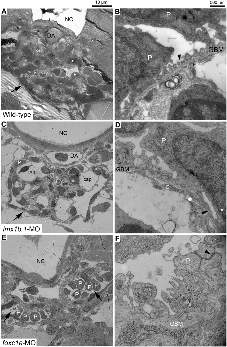
Figure 4.
Glomerular ultrastructures in lmx1b.1 and foxc1a morphants at 4 dpf. At low magnification, whole glomeruli (arrow) are exhibited (A, C, and E). Compared with wild-type (A), capillary lumens in the lmx1b.1 morphant are abnormally enlarged (C), and the foxc1a morphant shows that two glomeruli fail to merge at the midline and glomerular capillaries are missing (E). Detailed morphology of the glomerular filtration barrier is displayed at higher magnification (B, D, and F). In wild-type, fine foot processes, slit diaphragm (arrowhead) and fenestrated endothelium together with GBM are clearly visible (B). The lmx1b.1 morphant displays typical effacement (arrowhead), lack of endothelial fenestration, and a complete loss of the slit diaphragm (D). The foxc1a morphant shows aberrant podocyte foot processes on disorganized GBM and absence of endothelial cells. Normal cell-cell junctions by the slit diaphragm disappear and instead typical adherens junctions (arrowhead) occur between adjacent podocytes (F). cap, capillary; DA, dorsal aorta; E, endothelium; NC, notochord; P, podocyte. Magnification is shown by scale bars 10 µm and 500 nm.

