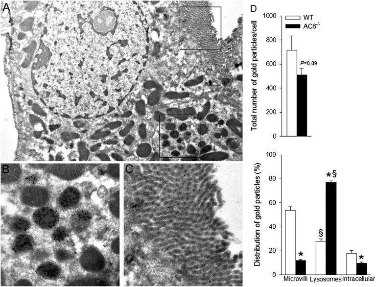
Figure 4.
The majority of Npt2a resides in lysosomes within proximal tubule cells of AC6−/− mice. (A) Low-magnification immunogold electron microscopy image showing the structure of the proximal tubule cell and the areas highlighted in B and C. At low magnification, gold particles can be observed in defined intracellular structures. (B) At higher magnification, gold particles representing Npt2a are abundantly observed in lysosomes. (C) Little Npt2a labeling was associated with the apical microvilli. Gold particles are 10 nm in diameter. (D) The distribution of colloidal gold particles representing Npt2a was counted within individual proximal tubule cells. Distribution of gold particles was subdivided into the apical microvilli, intracellular compartments, and lysosomes (n=4/genotype). *P<0.05 versus WT. §P<0.05 versus microvilli and intracellular same genotype.

