
Figure 8.
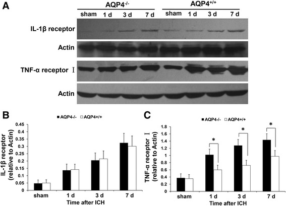
Expression of IL-1 β and TNF-α receptors in AQP4 −/− and AQP4 +/+ mice. It was revealed by Western blotting that higher levels of IL-1β and TNF-α receptors were detected in AQP4−/− mice than in AQP4+/+ mice at 1, 3, and 7 days after ICH (n =6, * P <0.05). (A) Western blotting images. (B) Semiquantitative analysis of IL-1β receptor. (C) Semiquantitative analysis of TNF-α receptor I. ICH, intracerebral hemorrhage.