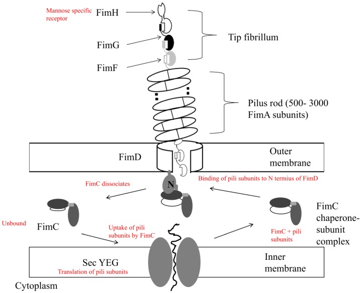
Figure 1.
The assembly of the type I pilus. The periplasmic protein FimC binds secreted pilus subunits, from the SecYEG translocon based in the internal membrane, to the periplasm. A process of accelerated subunit folding by FimC (periplasmic chaperone) occurs, followed by delivery to the usher outer assembly platform FimD, also performed by FimC. These FimC-subunit complexes are recognized and bind to the N-terminal domain of the usher: FimDN. Uncomplexed FimC is then released to the periplasm when subunits are assembled into the pilus. The tip of the pilus (fibrillum) consists of the protein adhesins FimF, FimG, and FimH, with FimA forming the bulk of the pilus rod. Adapted from Capitani, 2006 [37].

