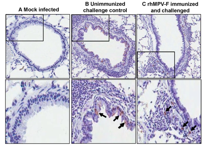
Figure 8.
Immunohistochemical (IHC) staining of lungs of cotton rats. (A) Lungs from mock infected cotton rats. (B) Lungs from unimmunized challenge control group. A large number of hMPV antigen-positive cells were detected at the luminal surfaces of bronchial epithelial cells. (C) Lungs from cotton rats immunized by rhMPV-F and challenged with the same virus. hMPV antigens were found inside the bronchial tissue, but not on the luminal surface of the bronchial epithelial cells.

