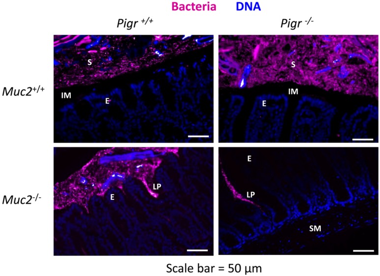
Figure 4.
Invasion of bacteria into the colonic lamina propria in the absence of Muc2. Freshly dissected colons from 6–8 week-old Muc2+/+Pigr+/+, Muc2−/−Pigr+/+, Muc2+/+Pigr−/− and Muc2−/−Pigr‑/− mice were preserved in Carnoy’s fixative and whole mounted to preserve the integrity of the mucus layer. Sections of colon tissue were stained by fluorescence in situ hybridization for bacterial 16S rRNA (magenta), then counterstained with DAPI to visualize cell nuclei (blue). Scale bar = 50 µM. E, enterocytes at the luminal surface; IM, inner mucus layer; LP, lamina propria; S, stool; SM, sub-mucosa. The inner mucus layer in Muc2+/+ mice is evidenced by the gap between the epithelial surface and bacteria present in the outer mucus layer and stool. Bacteria are in direct contact with the epithelial surface in Muc2−/− mice. The boundary between the outer mucus layer and stool cannot be distinguished with this staining method. Images are representative of multiple locations along the colon for at least three mice of each genotype.

