Figure 2.
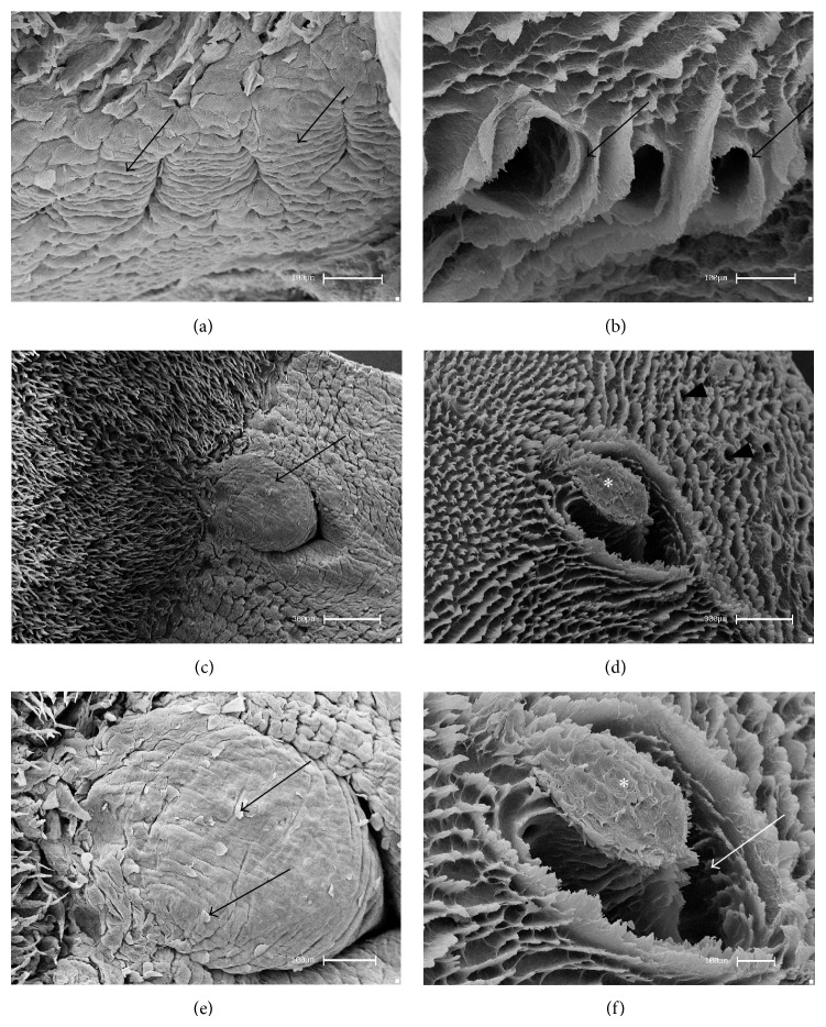
Scanning electron micrographs—foliate and vallate papillae of rats. (a) The foliate papillae are constituted by epithelial folds (arrows) and separated by parallel grooves. (b) After the removal of the epithelial tissue, the CTCs of the foliate papillae showed wide grooves limited by laminar projections (arrows). (c) The vallate papilla (arrow) is situated in the caudal region and (d) after the removal of the epithelium the general aspects of the CTCs (∗), delimited by CTCs of the filiform papillae and salivary glands ducts (arrowheads), were observed. (e) At higher magnification, squamous epithelium (arrows) in the surface was noted. (f) At higher magnification the constitution of the CTC by a thick bundle (∗) margined by a wide groove (arrow) was revealed. Bars: 100 μm (a, b, e, f) and 300 μm (c, d).

