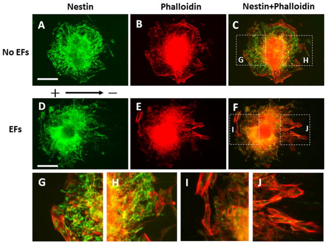
Fig. 3.
Orientation of the cells that migrated out of the embryoid bodies (EBs). The EBs were labelled with anti-nestin antibody (green) and Rhodamine Phalloidin (red). a–c The symmetrical distribution and random orientation of the cells that migrated out the EB without direct current electric fields (EFs) stimulation. g, h The magnified images show the dash line labelled areas in (c). d–f The asymmetrical distribution of cells that migrated out of the EB. On the cathode facing side (right), the cells aligned parallel to the field line of EFs. On the anode facing side (left) the cells oriented perpendicular to the field line of EFs. i, j Magnified images show the dash line labelled areas (f). Scale bar: 200 μm

