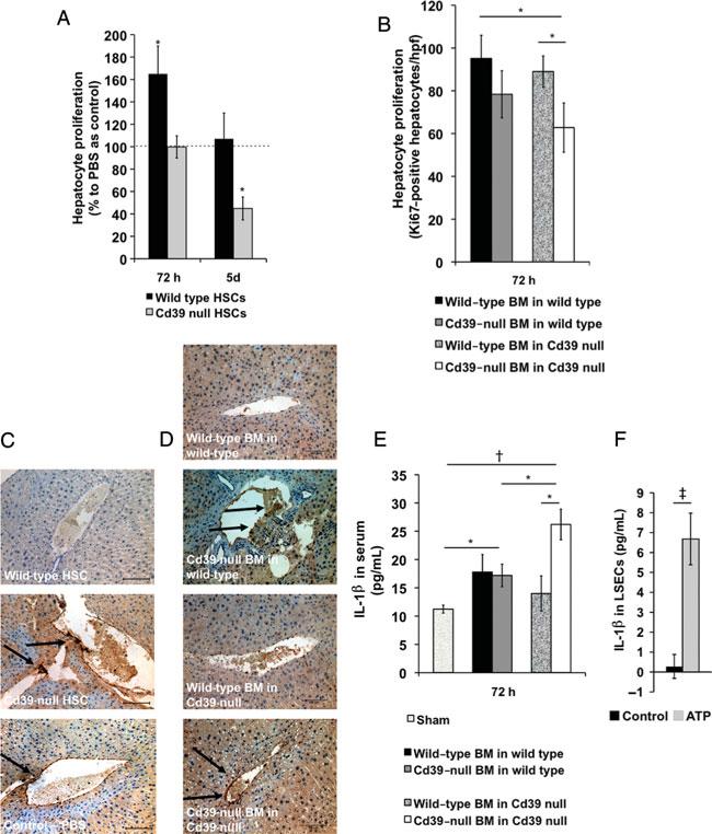
FIGURE 2.
Bone marrow- and HSC-dependent liver regeneration. A, Hepatocyte proliferation in regular wild-type mice receiving 3 105 wild-type or Cd39-null HSCs 24 hours×after 70% hepatectomy (% Ki67-positive hepatocyte nuclei/high-power field relative to wild-type mice receiving phosphate-buffered saline as control; n = 6–8). B, Hepatocyte proliferation in bone marrow-irradiated mice chimeric with wild-type or Cd39-null bone marrow 72 hours after 70% hepatectomy (Ki67-positive hepatocyte nuclei/hpf; 70% hepatectomy, n = 5–10). C, Representative immunostaining for vascular P-selectin in livers of regular wild-type mice receiving 3 × 10⁵ wild-type, Cd39-null HSCs or phosphate-buffered saline as control 24 hours after 70% hepatectomy (sham operation, n = 3; 70% hepatectomy, n = 6–8). Scale bars: 100 μm. D, Representative immunostaining for vascular P-selectin in livers of bone marrow-irradiated mice chimeric with wild-type or Cd39-null bone marrow 72 hours after 70% hepatectomy (n 6–10). Scale bars: 100 μm. E, interleukin-1β=plasma concentration (pg/mL) in bone marrow-irradiated mice chimeric with wild-type or Cd39-null bone marrow at 72 hours (sham operation, n = 3; 70% hepatectomy, n = 6–8). F, interleukin-1β concentrations (pg/mL) in liver sinusoidal endothelial cells, cultured for 5 hours with/without preincubation with adenosine triphosphate (5 mM) for 20 minutes. Error bars represent standard error of mean. *P < 0.05, †P < 0.01, ‡P < 0.001.

