Fig. 4.

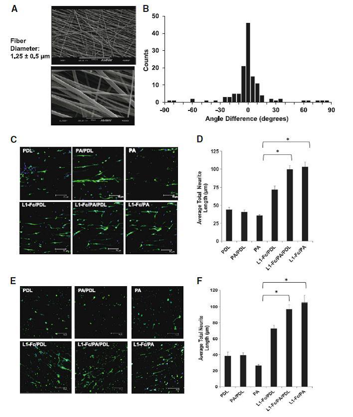
Protein A-presented L1-Fc increases neurite outgrowth in aligned, polymeric scaffolds. a Morphology and fiber diameter of aligned scaffolds that were fabricated by electrospinning. b Histogram depicting the degree of fiber alignment of the scaffolds. c Spinal cord neurons (SCNs) were plated onto scaffolds treated with L1-Fc presented from PDL, PA/PDL, or PA alone, as well as control surfaces that did not contain L1-Fc. PA presented L1-Fc results in longer neurite lengths when cultured on aligned scaffolds. Neurons cultured on 10 μg/ml L1-Fc passively adsorbed onto the fibers extended longer neurites than on the PDL control. The three-dimensional scaffold results are similar to those observed on the polymer thin films. d Quantification of the average total neurite lengths of SCNs cultured on the different L1-Fc presentations showed L1-Fc/PDL resulted in SCNs with shorter neurites compared to L1-Fc/PA/PDL and L1-Fc/PA, suggesting that the manner in which L1-Fc is presented on nanofibrous scaffolds influences its efficacy. (e, f) Cerebellar neurons (CNs) were plated onto scaffolds pretreated with different L1-Fc presentations and controls as mentioned above. PA presented L1-Fc resulted in CNs that were more spread and grew longer along the fibers compared to CNs cultured on L1-Fc adsorbed onto PDL. The three-dimensional scaffold results correlate to the results observed on the polymer thin films. Scale bar 75 μm. PDL poly-d-lysine, PA protein A (* denotes p < 0.05)
