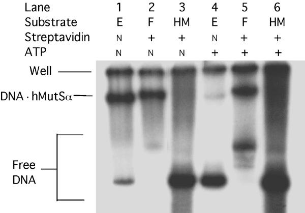
Figure 3.

ATP-resistant retention of hMutSα on end-blocked mismatched DNA. The indicated 0.5-kb DNA fragments (6 fmol) cleaved from biotinylated, mismatched-DNA substrate (E) (Figure 1A) or nonderivatized homoduplex DNA (HM) were purified and endlabeled, and incubated first with streptavidin in binding buffer (to convert substrate (E) to substrate (F)), and then with purified hMutSα (0.25 pmol) plus 125 ng nonspecific competitor DNA for 10 min, as described under ‘Materials and methods'. After addition of ATP to 200 μM, where indicated, incubations were continued for an additional 5 min. Mixtures were separated by electrophoresis in 5% nondenaturing polyacrylamide gels (acrylamide to bisacrylamide ratio of 37.5:1), which were dried and analyzed by phosphorimaging. The fastest-moving DNA bands (lanes 1, 3, 4, 6) correspond to free DNA fragments (not bound to MutSα) from substrates (E) or (HM). The slower-moving faint free-DNA band in lane 2 corresponds to the small amount of substrate (F) with both streptavidin complexes but not bound to hMutSα, as do the more intense free DNA bands in lane 5. The two faint bands moving slightly faster in lane 5 presumably correspond to fragments with one complex, either 150 bp from one end or 20 bp from the other end—locations expected to affect mobility differentially. ATP would be expected to cause hMutSα to slide off the free ends of both the latter DNA species. Intensities of the indicated slowest-moving bands, corresponding to DNA hMutSα complexes, were compared to the total of bound plus nonbound free DNA, excluding DNA not fully complexed with streptavidin (faint bands), and disregarding DNA trapped in the wells (‘Well'). Bound fractions were 84 and 90%, respectively, for biotinylated-only (lane 1) and streptavidin-complexed (lane 2) DNA in the absence of ATP, but 12 and 51%, respectively, for these substrates in the presence of ATP (lanes 4 and 5).
