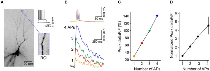
Figure 3.

Brief trains of back-propagating APs cause linear increases in Ca2+ in L3 PC distal apical dendrites. (A) L3 PC (identified by morphology and intrinsic electrical properties) filled with the calcium-sensitive dye OGB1-AM. Blue box, distal region of interest (ROI) used for measurements of Ca2+ transients. (B) Trains of 1–4 APs (50 Hz) evoked via somatic current injection (top traces) elicit Ca2+ transients (bottom traces, deltaF/F) in the ROI from cell in (A). (C) The relationship between peak dF/F and number of APs is linear in this range. (D) Summary of results (n = 8 cells, error bar = ±SEM) shows linear relationship between Ca2+ and number of APs.
