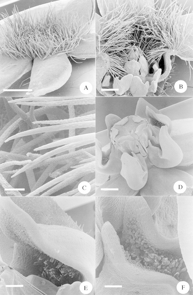
Fig. 6. A, Scanning electron micrograph (SEM) of a whole flower of Sisyranthus trichostomus. Scale bar = 1·0 mm. B, SEM of a half flower of Sisyranthus trichostomus. Note the hairs blocking the corolla tube arching down into the nectar‐holding corona cups. Scale bar = 0·5 mm. C, SEM close‐up of the hairs blocking the corolla tube of Sisyranthus trichostomus, showing the parallel ridges running along the main axis of each hair. Scale bar = 50 µm. D, SEM of a whole flower of Asclepias cucullata. Scale bar = 1·0 mm. E, SEM close‐up of the portion of the nectar cup distal to the gynostegium of Asclepias cucullata. Note the dense papillae within the cup. Scale bar = 250 µm. F, SEM close‐up of the portion of the nectar cup proximal to the gynostegium of Asclepias cucullata. Note the dense papillae within the cup. Scale bar = 150 µm.
