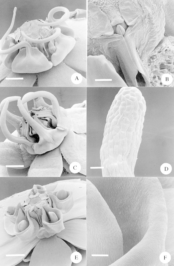
Fig. 8. A, SEM of whole flower of Miraglossum verticillare. Note that the spiral arms of the two distant corona elements were lost during specimen processing. Scale bar = 1·0 mm. B, SEM close‐up of the guide rails of Miraglossum verticillare, with the pollinarium in situ. To the left is the edge of an adjacent corona element; the one to the right has been removed. Scale bar = 250 µm. C, SEM of whole flower of Miraglossum pilosum. Note that the filiform corona elements are usually straight and were deformed during specimen processing. Scale bar = 1·0 mm. D, SEM close‐up of the terminal end of a filiform corona element of Miraglossum pilosum. Scale bar = 50 µm. E, SEM of whole flower of Aspidonepsis diploglossa. Scale bar = 1 mm. F, SEM close‐up of the inner wall of the nectar cup of Aspidonepsis diploglossa. To the left is the ‘tongue’ situated within the cup. Scale bar = 100 µm.
