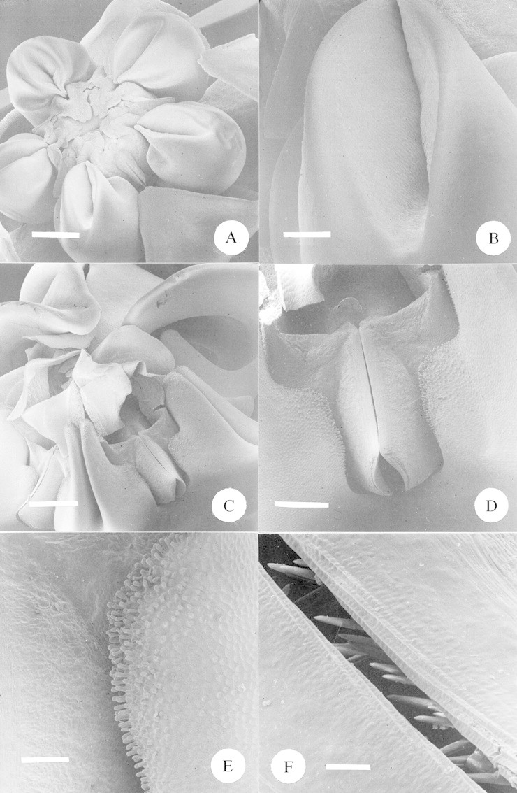
Fig. 9. A, SEM of a whole flower of Asclepias woodii. Scale bar = 1·0 mm. B, SEM close‐up of the inner surface of the nectar ‘cup’ of Asclepias woodii. Scale bar = 0·25 mm. C, SEM of the central portion of the large flower of Pachycarpus natalensis. The over‐arching portions of the three closest corona lobes have been removed to facilitate a view of the gynostegium. Scale bar = 1·0 mm. D, SEM close‐up of guide rails of Pachycarpus natalensis. The adjacent corona lobes fuse with the gynostegium at either side. Scale bar = 0·5 mm. E, SEM close‐up of Pachycarpus natalensis, showing the point at which the gynostegium (to the left) meets with the corona (to the right). Note the densely papillate epidermis of the corona. Scale bar = 150 µm. F, SEM close‐up of the guide rails of Pachycarpus natalensis. Note the upward‐pointing ‘teeth’ within the guide rails. Scale bar = 20 µm.
