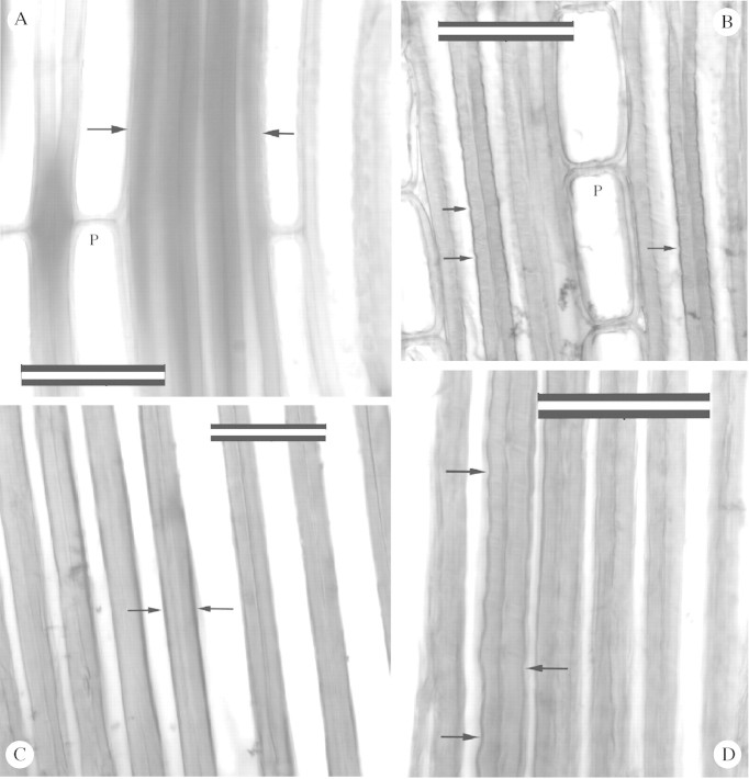
Fig. 3. Fibres of Quercus sp. and latewood tracheids of Metasequoia as seen in radial longitudinal views. A, Non‐compressed (normal) wood of Quercus. Arrows indicate fibres that have smooth walls. P, Parenchyma cell. B, Quercus wood that has been compressed to approx. 85 % of its original length. Arrows point to corrugated walls which also display slip planes (light lines). P, Shortened parenchyma cell. C, Latewood tracheids from the outermost ring (26) of PNJ Metasequoia tree. Arrows point to tracheids where plastic deformation is minimal or absent. D, Latewood tracheids of ring 14 of PNJ Metasequoia. Arrows point to corrugations in tracheid walls, and slip planes are evident. Bars = 50 µm.

An official website of the United States government
Here's how you know
Official websites use .gov
A
.gov website belongs to an official
government organization in the United States.
Secure .gov websites use HTTPS
A lock (
) or https:// means you've safely
connected to the .gov website. Share sensitive
information only on official, secure websites.
