
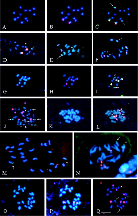
Fig. 1. Fluorescent in situ hybridization with 5S (green) and 45S (red or yellow) rDNA probes, in Passiflora species with x = 6, x = 10 or x = 12. A–C, P. capsularis. Note chromosomes stained with DAPI in A, 45S rDNA sites in B and a pair of 5S rDNA sites in C (arrows). D, P. rubra. Arrows indicate 5S rDNA. E, P. morifolia. Arrows indicate 5S rDNA. F, P. tricuspis. Note a longer pair of chromosomes with terminal regions characteristically less condensed, two pairs with proximal blocks of 45S rDNA, with one of the chromosomes being distended (arrowhead), and a pair with sub‐terminal 5S rDNA (arrows). G–I, P. misera 2x. Note the terminal DAPI+ bands in almost all chromosomes in G, 45S rDNA sites apparently sub‐terminal in one pair of chromosomes and proximal in the other one in H, and 5S rDNA sites in I (arrows). J, P. misera 6x. Broken arrows and arrowhead indicate small 45S rDNA sites. K and L, P. suberosa. Note that three of the five pairs of 45S rDNA sites are located in the smallest chromosomes, two of which also have 5S rDNA (arrows). M, P. haematostigma. Arrows indicate terminal 5S rDNA sites. N, P. pentagona. Arrows indicate 5S rDNA sites. O–Q, P. foetida. Note the secondary proximal constrictions in three chromosome pairs in O, three pairs of 45S rDNA proximal sites in P, and two pairs of 5S rDNA sub‐terminal sites in Q (arrows). All chromosomes were counterstained with DAPI (blue). Bar in Q represents 5 µm.