
Figure 5.
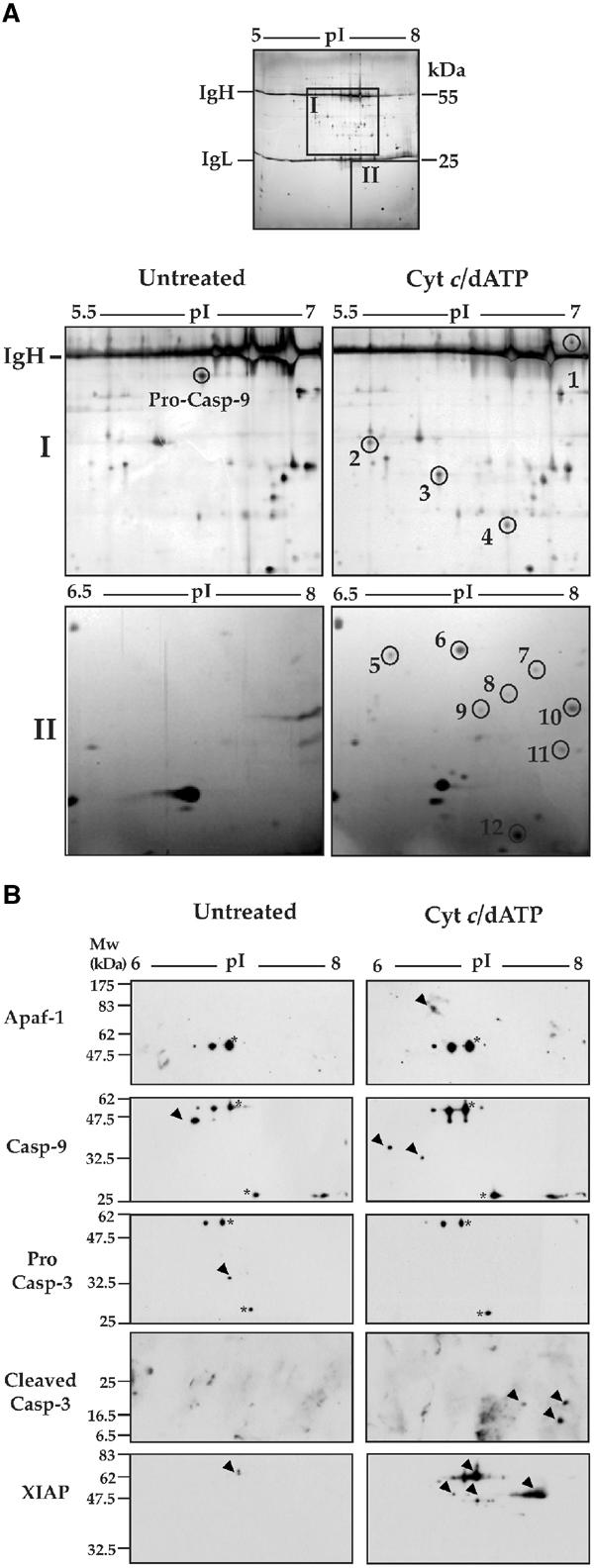
Analysis of native Apaf-1 apoptosomes by 2D-PAGE. (A) Caspase-9 complexes, isolated from 2 ml Jurkat cell-free reactions incubated for 15 min at 37°C in the presence or absence of 50 μg/ml cytochrome c/1 mM dATP, were analyzed by 2D gel electrophoresis (first dimension: pH 5–8, second dimension: 12% SDS–PAGE). Top: A representative silver-stained preparative gel is shown. Heavy and light chains of the immunoprecipitating antibody are indicated. Bottom: enlarged areas containing proteins that differentially immunoprecipitate with caspase-9 (areas I and II in the gel above) are shown. (B) Caspase-9 complexes were isolated from Jurkat cell-free extracts incubated for 15 min at 37°C in the presence or absence of 50 μg/ml cytochrome c/1 mM dATP. Proteins co-precipitating with caspase-9 were analyzed by 2D gel electrophoresis (pH 5–8 first dimension and 12% SDS–PAGE second dimension), followed by immunoblotting with the indicated antibodies. Spots corresponding to immunoglobulin heavy and light chains are indicated by asterisks.