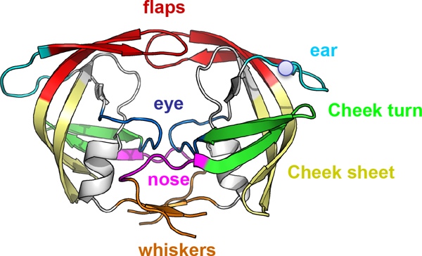
Figure 1.

Topology of the HIV-1 protease. Topology of HIV-1 protease colored according to the convention proposed in Perryman et al, 2003. In red the flap domain (residues 43-58); in cyan the ears (35-42); in green the cheek turns (11-22); in yellow the cheek sheets (59-75); in blue the eyes (23-30); in magenta the nose (6-10) and in orange the whiskers (1-5 and 95-99). The 36th residue is represented by a grey sphere.
