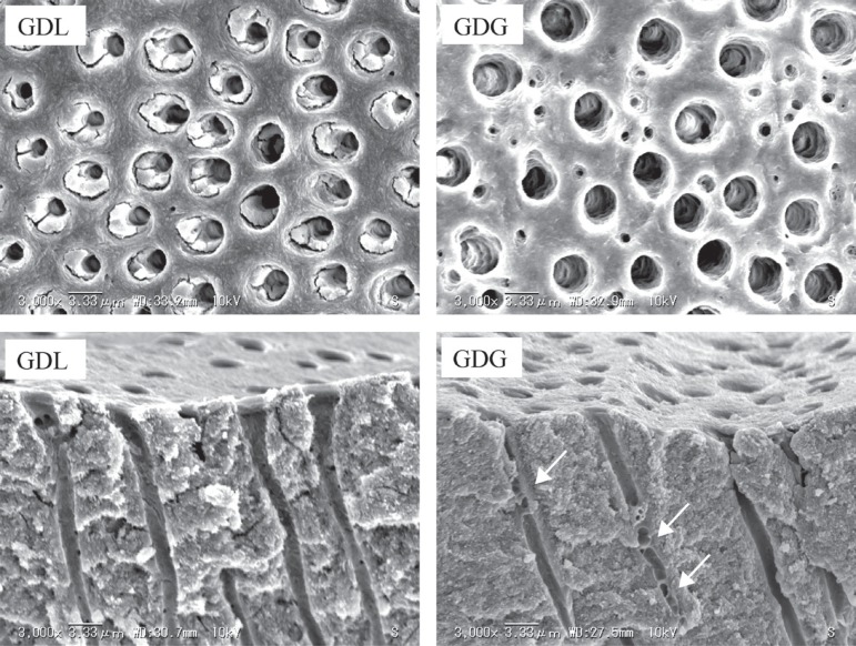
Figure 5.
Representative scanning electron microscope micrographs (3,000x) of GDL and GDG treated dentin disc surfaces and of samples fractured perpendicular through the desensitizer-treated surfaces after rinsing with water. The morphologies of the treated surfaces are similar; no remnants of the thickening agent of GDG on free surface or inside tubules. In two of the longitudinally exposed tubules of GDG transverse septa are displayed (arrows)

