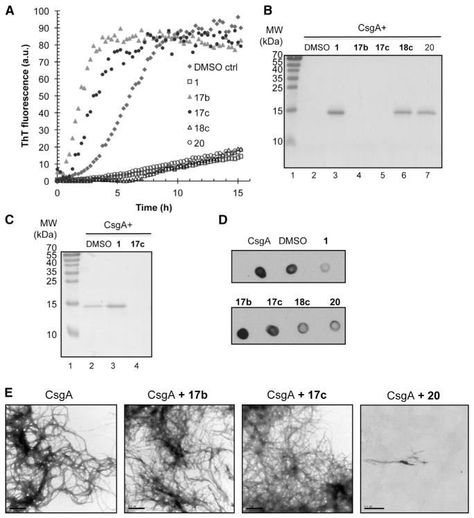
Figure 4. In Vitro Effect of Accelerating Compounds on CsgA Polymerization.
(A) ThT fluorescence of freshly purified 5 μM CsgA with or without 50 μM compound. The lag phase of the fluorescence curve as a consequence of ThT binding to polymerizing CsgA was shortened when accelerating compounds 17b and 17c were present.
(B) SDS-PAGE of CsgA incubated overnight with or without compound. No soluble CsgA was detected after incubation with accelerators 17b and 17c (lanes 4 and 5, respectively).
(C) SDS-PAGE of CsgA incubated for 8h with or without compound. Incubation with the accelerating compound 17c results in a SDS-insoluble amyloid after 8 hr (lane 4) whereas pure CsgA is still partly soluble (lane 2). In the presence of the inhibitory compound 1, CsgA is soluble (lane 3).
(D) CsgA incubated overnight with compounds was spotted onto a nitrocellulose membrane and probed with the Tessier gammabody grafted with the CsgA R1 sequence that detects CsgA amyloid structure.
(E) Transmission electron microscopy of CsgA. Negative staining of freshly purified CsgA incubated for 6 hr (left). Freshly purified CsgA incubated for 6h with accelerators 17b or 17c appeared similar to CsgA alone. Fibers were not detectable when CsgA was incubation with the chemically similar, yet functionally inhibitory, compound 20. Scale bars represent 0.5 μM.
See also Figure S1.

