
Figure 1.
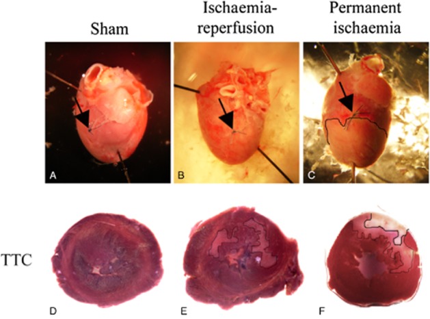
Montage demonstrating representative (A) sham-operated, (B) ischaemia-reperfused and (C) permanent ischaemic hearts. Upper panel shows hearts with the surgical ligature (arrows) and in the lower panel, TTC staining of heart slices. The thin line (in D, E) outlines areas with diffuse ischaemic appearance and the thicker line (only in permanent ischaemic hearts; F) with clear infarction.