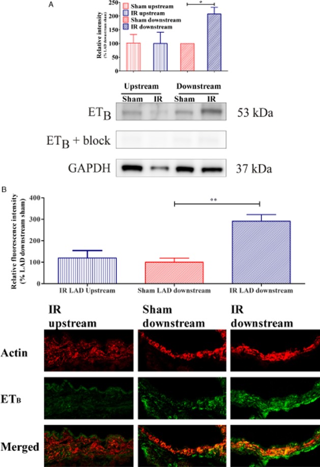
Figure 4.

(A) Western blot of three coronary artery homogenates pooled from IR- (n = 10) and sham- (n = 9) operated rats. Results revealed endothelin ETB receptor band at approximately 50 kDa. Quantification of ETB immunoreactivity normalized to GAPDH revealed increased ETB receptor protein levels in downstream LAD segments after IR. Results are expressed as mean ± SEM of three different homogenates where mean of (ETB immunoreactivity)/(GAPDH immunoreactivity) from sham LAD downstream was set to 100%. *P < 0.05, comparing LAD downstream IR versus LAD downstream sham; Student's t-test. (B) The fluorescence intensity of each group is given as the percentage fluorescence of IR LAD downstream (n = 5) or IR LAD upstream (n = 3) group compared with the sham LAD downstream (n = 3) groups, where the LAD downstream group was set to 100%, and the mean value for each of the other groups was used for comparison. Confocal images showing representative examples of immunofluorescence staining of rat coronary arteries (n = 3–5) of actin (upper row), ETB receptor (middle row), and merged pictures that show the location of endothelin ETB receptors in VSMC (bottom row). **P < 0.01, comparing LAD downstream IR versus LAD downstream sham; Student's t-test.
