FIGURE 2.

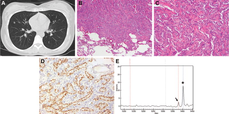
Case 5. A, Suspicious density seen on CT scan in the right middle lobe of the lung at the time of diagnosis of osteosarcoma of the humerus. B and C, Invasive adenocarcinoma of the right middle lobe of the lung. Note the presence of invasive glands with fibrotic stroma and sharp demarcation between the tumor cells and the normal pulmonary parenchyma (hematoxylin and eosin; original magnifications, 10× and 40×). D, Immunohistochemical stain for TTF-1. This photomicrograph shows that the invasive glands are positive for TTF-1, thus confirming a pulmonary origin for this adenocarcinoma. E, Mass spectrometry-based genotyping for EGFR exon 21 L858R mutation on Sequenom platform confirming the presence of L858R mutation. The germline peak is indicated by an asterisk and the mutant peak by an arrow. CT, computed tomography; TTF, thyroid transcription factor-1.
