
Figure 2.
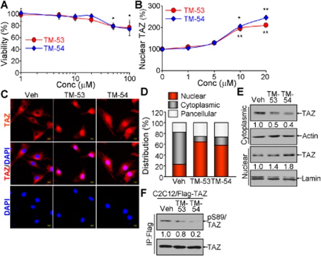
Enhancement of TAZ nuclear translocation by TM-53 and TM-54. (A) C2C12 cells were treated with TM-53 and TM-54 (1, 5, 10, 20, 50 and 100 μM) for 24 h and subjected to the cytotoxicity assay. Cell viability was calculated by measuring optical density at 450 nm after staining and is given as mean ± SEM for three experiments. The viability of cells treated with vehicle was set as 100%. (B) Cells were treated with TM-53 and TM-54 (0, 1, 5, 10 and 20 μM) and stained with anti-TAZ antibody. The location of the nuclei was indicated by DAPI staining. Nuclear TAZ was calculated by dividing nuclear TAZ-expressing cells by the total number of TAZ-expressing cells (100 cells for each counting, five times). Nuclear TAZ expression in vehicle-treated cells was regarded as 100% and is expressed as mean ± SEM. (C, D) C2C12 cells were treated with 10 μM TM-53 and TM-54 for 24 h. Cells were subjected to immunofluorescence staining with anti-TAZ antibody. Representative images of three independent experiments are shown (C). Percentages of nuclear, cytoplasmic and pan-cellular TAZ distribution were calculated by counting 80–150 cells in multiple fields of view (n = 5 per sample) (D). (E, F) C2C12 cells stably expressing Flag-TAZ were incubated with TM-53 (10 μM) and TM-54 (10 μM) for 24 h. Cells were harvested and subjected to fractionation for nuclear and cytoplasmic protein, followed by immunoblotting analysis (E). Total protein extract was used for immunoprecipitation with anti-Flag antibody and subsequently analysed by SDS-PAGE and immunoblotting with anti-pS89 TAZ antibody.