
Figure 6.
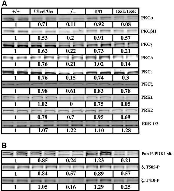
Immunoblotting of PKC isoforms in ES cells. The indicated ES cells cultured in the presence of 10% serum were lysed and aliquots (20 μg of protein) were subjected to SDS–polyacrylamide gel electrophoresis and immunoblotted with the indicated total PKC isoform antibodies (A) or phosphospecific T-loop PKC antibodies (B). Quantitation of the immunoblots, indicated immediately below each blot, was performed using LI-COR Odyssey infrared imaging system as described in Materials and methods. The value of expression of each PKC isoform detected in PDK1(+/+) cells is taken as 1.0 and the expression of the isoforms in other cells expressed relative to this. As a control, the cell lysates were immunoblotted for ERK1 and ERK2 whose levels are not regulated by PDK1. Similar results were obtained in at least three independent experiments.