
Figure 9.
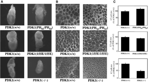
The PDK1(PHKI/PHKI) and PDK1(155E/155E) cells are of normal size. (A) The indicated E7.0 PDK1 knockin or knockout embryos were dissected in PBS and imaged on a Leica M275 microscope and whole-mount photographs were taken. Embryos are shown at the same magnification. In each case, the mutant PDK1 embryos are compared to their wild-type littermates. (B) Representative images of the indicated E7.0 embryonic endoderm cells stained with the lipid dye DilC16(3) using a Zeiss LSM510 microscope. (C) The size of the E7.0 embryonic endoderm cells was quantitated as described in Materials and methods. Three embryos of each genotype were analysed, with 120 cells in each embryo being measured and P<0.0004.