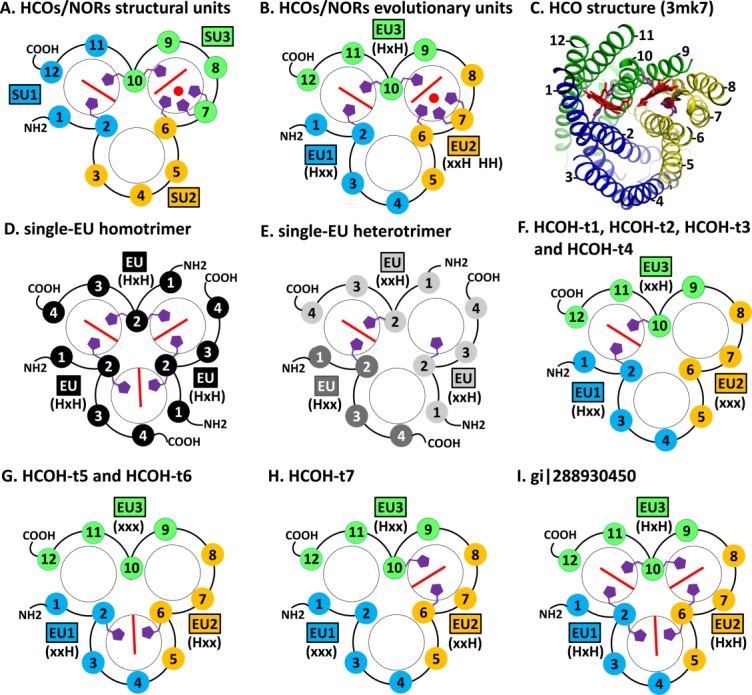
Figure 1.

TMH topology diagrams of HCOs/NORs and HCOH proteins. TMHs are shown in numbered circles. TMHs in the same structure unit (marked by SU) or the same evolutionary unit (marked by EU) are filled with the same color. The histidine patterns of the EUs are shown in parentheses. Hemes are shown as red lines. Copper or non-heme irons in HCOs/NORs are shown as red spheres. Conserved histidine sidechains are shown in purple. N- and C-termini of each modeled protein are marked by NH2 and COOH, respectively. (A). Previously defined structural units of HCOs/NORs. (B). Newly defined EUs of HCOs/NORs. (C). Structure of a C-type HCO catalytic subunit (pdb: 3mk7). (D). Model of homotrimer for single-EU proteins with the HxH motif. (E). Model of heterotrimer for single-EU proteins with xxH and Hxx motifs. (F). Model for HCOH-t1, HCOH-t2, HCOH-t3 and HCOH-t4. (G). Model for HCOH-t5 and HCOH-t6. (H). Model for HCOH-t7. (I). Model for the protein (gi|288930450) with the HxH motif in all three EUs.
