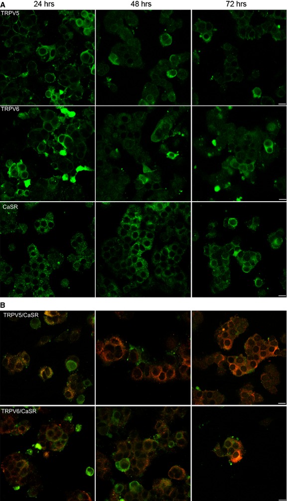
Figure 5.

Confocal images of primary human parathyroid cells maintained at different times of incubation. (A) It shows the expression of TRPV5, TRPV6 and CASR (green), respectively, at 24, 48 and 72 hrs. (B) It shows the co-localization between TRPV5, TRPV6 (green) and CASR (red). The immunofluorescence staining images were detected by a confocal laser scanning microscope (Leica TCS-SP5) by using 40× zoom 1 oil objective with 1.45 NA and a recommended pinhole size of less than 1.0 μm; scale bars: 10 μm.
