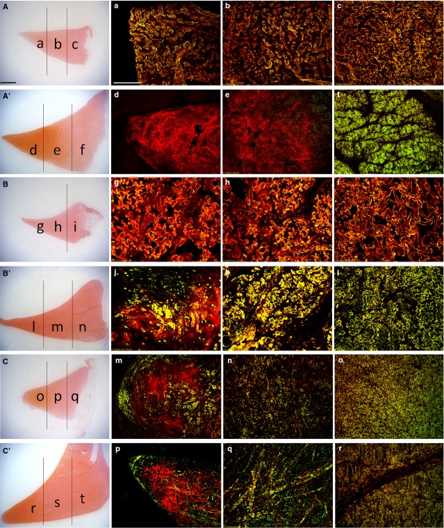
Figure 3.
Immunofluorescent staining of collagen 1 and 2 on traversal sections: medial anterior horn of the young (a–c) and adult (d–f) meniscus; medial body of the young (g–i) and adult (l–n) meniscus; medial posterior horn of the young (o–q) and adult (r–t) meniscus. Collagen 1 is in green, collagen 2 is in red, superimposition appears in yellow colour (various degrees). Scale bar 200 μm. For comparison, two representative pictures of the young (A–C) and adult (A’, B’, C’) anterior horns, body and posterior horn at low magnification, stained for SAFRANIN-O. Scale bar 2000 μm. (a) Inner zone, anterior horn young meniscus. (b) Intermediate zone, anterior horn, young meniscus. (c) Outer zone, anterior horn, young meniscus. (d) Inner zone, anterior horn, adult meniscus. (e) Intermediate zone, anterior horn adult meniscus, (f) Outer zone, anterior horn, adult meniscus. (g) Inner zone, body, young meniscus. (h) Intermediate zone, body, young meniscus. (i) Outer zone, body, young meniscus. (j) Inner zone, body, adult meniscus. (k) Intermediate zone, body, adult meniscus. (l) Outer zone, body, adult meniscus, (m) Inner zone, posterior horn young meniscus. (n) Intermediate zone, posterior horn, young meniscus. (o) Outer zone, posterior horn, young meniscus. (p) Inner zone, posterior horn, adult meniscus. (q) Intermediate zone, posterior horn adult meniscus. (r) Outer zone, posterior horn, adult meniscus.

