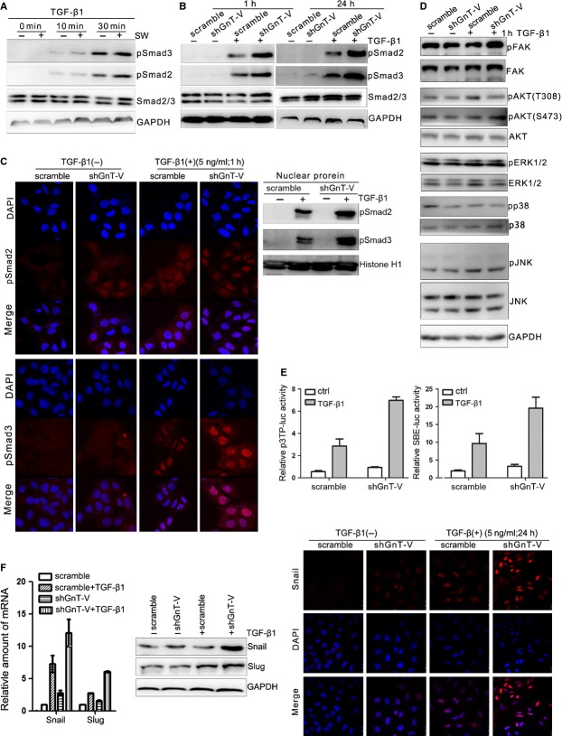
Figure 6.
Inhibition of β1,6-GlcNAc branched N-glycans' formation through swainsonine treatment or GnT-V knockdown in lung cancer cells enhances the activation of TGF-β/Smads signalling. (A) Swainsonine treatment enhances TGF-β1-mediated Smad2 and Smad3 phosphorylation in A549 cells, as determined the Smad2 and Smad3 proteins, and their phosphorylation levels (pSmad2, pSmad3) by western blot. A549 cells were pre-treated with or without swainsonine (1 μg/ml) for 24 hrs, before exposure to TGF-β1 (5 ng/ml) for indicated time periods. (B) Knockdown of GnT-V enhances TGF-β1-induced Smad2 and Smad3 phosphorylation in A549 cells. The phosphorylation and total protein levels of Smad2/Smad3 were determined by western blot. Scramble and shGnT-V A549 cells were exposed to TGF-β1 (5 ng/ml) for 1 or 24 hrs. (C) Knockdown of GnT-V enhances TGF-β1-induced nuclear translocation of pSmad2 and pSmad3 in A549 cells, as determined by immunofluorescence staining (left) and nuclear protein immunoblotting (right). Scramble and shGnT-V A549 cells were exposed to TGF-β1 (5 ng/ml) for 1 hr. (D) Knockdown of GnT-V does not alter TGF-β-non-Smad signalling in A549 cells. Scramble and shGnT-V A549 cells were exposed to TGF-β1 (5 ng/ml) for 1 hr, and the phosphorylation and total protein levels of signalling molecules (FAK, AKT, ERK, p38 and JNK) in TGF-β-non-Smad pathway were determined by western blot. GAPDH was used as loading control. (E) Knockdown of GnT-V increases TGF-β1-induced Smad2/Smad3 transcriptional reporter activity in A549 cells, as determined by a dual-luciferase assay. Scramble and shGnT-V A549 cells were transiently transfected by Smad2/4 driven-3TP-luciferase and Smad3/4 driven-(SBE)4-luciferase. After transfection, cells were treated with or without TGF-β1 (5 ng/ml) for another 24 hrs, then dual-luciferase assay was performed. (F) Knockdown of GnT-V increases TGF-β1-induced Snail/Slug expression and nuclear translocation in A549 cells, as determined by qRT-PCR (left) and western blot (middle), and immunofluorescence staining (right) of A549 cells' exposure to TGF-β1 (5 ng/ml) for 24 hrs.

