
Figure 2.
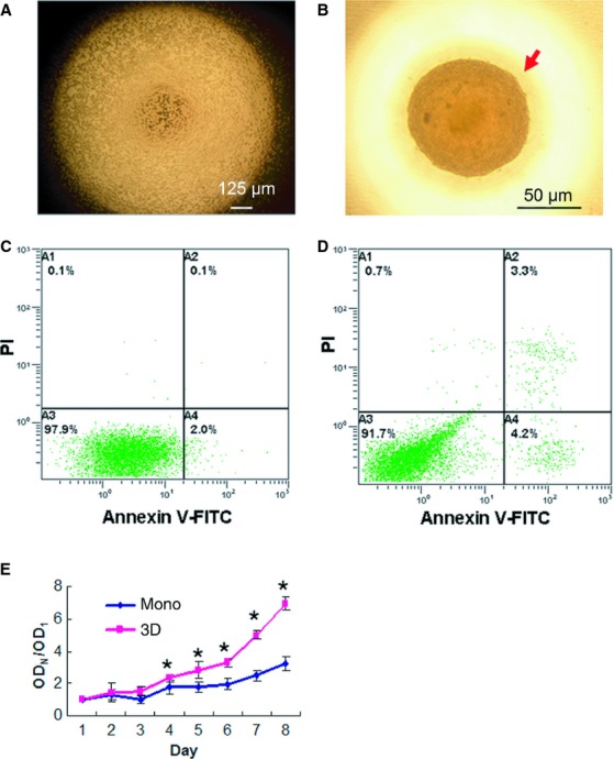
Viability and proliferation of spheroid hMSCs. 3 × 10⁴ hMSCs were suspended in 10% FBS in a hanging drop and incubated for 36 hrs to form spheroids. (A) It shows a microscopic image of the hanging drop at 0 hr. (B) A representative sphere of the hanging drop at 36 hrs was photographed under microscope. The spheroids were then transferred to suspension culture and incubated for 24 hrs. Cells derived from the spheroids were subjected to Annexin V/PI analysis by flow cytometry (D). hMSCs that were in the same passage but cultured in monolayer were used as a control (C). (E) MTT assay. Single hMSCs derived from spheroids (3D) or monolayer (mono) were seeded into tissue culture plates, incubated and subjected to MTT analysis. Spheroid hMSCs showed greater proliferating rates. Triplet wells were used for the assay, and the experiment was repeated three times with similar results (*P < 0.05). ODN represents OD values in corresponding days, ODN/OD1 represents the ratio of ODN to OD1 (the OD value in the first day).