
Figure 5.
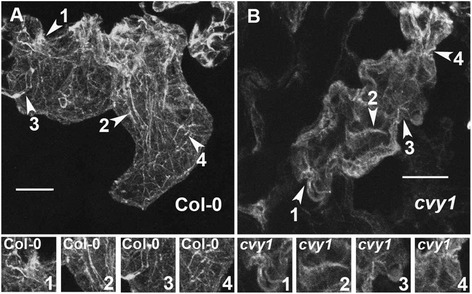
Knockout cvy1 null mutant displays reduced and disorganized actin bundles. (A and B) Actin organization in wild-type and curvy1 pavement cells was visualized using fluorescent phalloidin as previously described [2]. Depicted regions (arrow heads with numbers) of WT and curvy1 pavement cells were magnified in the bottom panels to display the actin bundles in respective genotype backgrounds. Bars = 10 μm.