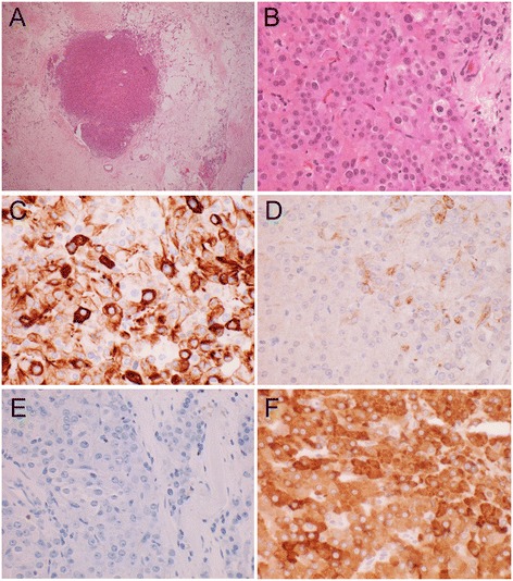
Figure 4.

Immunohistochemical staining of SRLC tumor. A) Tumor nodule, (4×) B) Tumor section H&E stained (40×) Leydig cells and scattered Sertoli cells. C) Strong to weak CAM 5.2 staining 40× D) Weak and specific Keratin A/E staining 40× E) Epithelial Membrane Antigen (EMA) Negative. Sertoli and Leydig cells are both EMA negative, 40× F) Inhibin Positive, 40×. Both Sertoli and Leydig cells typically show a Calretinin+, CD99+, Inhibin + phenotype.
