
Figure 2. NMR structures of WT human insulin and [GlnB22]-insulin.
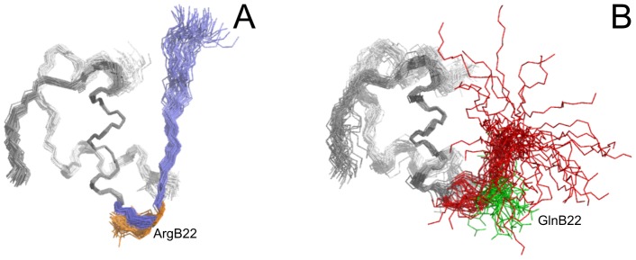
(A) Structures of WT human insulin are represented by a best-fit superposition of the protein backbone for 50 converged structures, with the positions of arginine B22 side chains colored in orange. B20–B30 protein backbone in WT human insulin is colored in blue. (B) Structures of [GlnB22]-insulin are represented by a best-fit superposition of the protein backbone for 40 converged structures, with the positions of glutamine B22 side chains colored in green. B20–B30 protein backbone in [GlnB22]-insulin is colored in red. A1–A21 and B1-B19 protein backbones in both insulins are colored in grey.