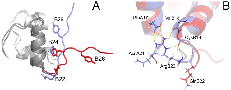
Figure 3. Overlay of representative NMR structures of WT human insulin and [GlnB22]-insulin.

(A) Overlay of the representative structures of WT human insulin (blue) and [GlnB22]-insulin (red). (B) Detailed view of an overlay of the B20–B23 β-turn and its surrounding area in the representative structures of WT human insulin (blue) and [GlnB22]-insulin (red). The network of hydrogen bonds stabilizing the B20–B23 β-turn in WT human insulin is highlighted by dashed yellow lines.
