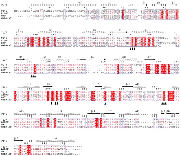
Figure 3. Structure-based sequence alignment of YgjG, AcOAT (PDB code 1VEF), OAT (PDB code 2OAT), and GABA-AT (PDB code 1SFF).
The amino acid numbering at the top of the alignment is for E. coli YgjG. The residues involved in the PLP-binding sites are indicated by black triangles, and the conserved lysine residues are indicated by a blue triangle. The white letters on red background indicate fully conserved residues, while the red letters on white background indicate partially conserved residues. The secondary structural elements depicted at the top of the alignment correspond to those in YgjG. The structure-based sequence alignment figure was generated using ESPript [27].

