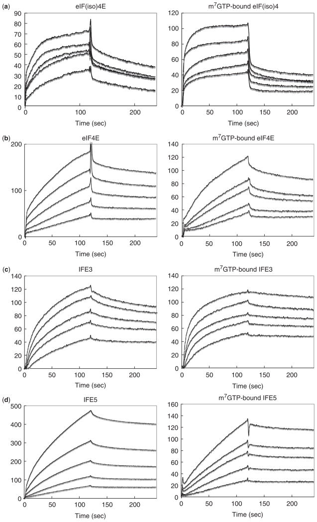
Fig. 1. SPR sensorgrams for the interaction of immobilized TuMV VPg with (a) A. thaliana eIFiso4E, (b) human eIF4E, (c) C. elegans IFE-3, and (d) C. elegans IFE-5.
The left and right parts correspond to the m7GTP-free and -bound eIF4E orthologues, respectively. Each SPR signal reflects the response after buffer containing the eIF4E orthologue (analyte) begins to flow over a CM dextran sensor chip to which the VPg is coupled through amino groups. The concentrations of analytes, corresponding to curves of increasing RU, are: 5.0–20.0 μM and 1.3–5.0 μM for m7GTP-free and -bound forms for (a), 0.2–2.5 μM and 1.3–30.0 μM for (b), 0.6–7.5 μM and 0.1–0.9 μM for (c) and 0.3–5.0 μM and 0.6–10.0 μM for (d). Each sensorgram consist of a formation portion (injection of analyte) and a dissociation portion (injection of buffer alone).

